Feline Mast Cell Tumors: Cutaneous Masses, Splenic Disease and Treatment
Feline mast cell tumors (MCTs or mastocytomas) represent the second most common skin neoplasm in cats and a prominent visceral malignancy, arising from the neoplastic transformation of granule-laden tissue mast cells. Unlike the aggressive biological behavior frequently seen in dogs, feline cutaneous mastocytomas typically behave in a benign manner and are curable with surgical excision. Conversely, visceral mast cell disease—most notably primary splenic mastocytoma—induces marked splenomegaly, systemic illness driven by histamine degranulation, and warrants urgent splenectomy.
In this guide
Cutaneous vs. Visceral Presentations
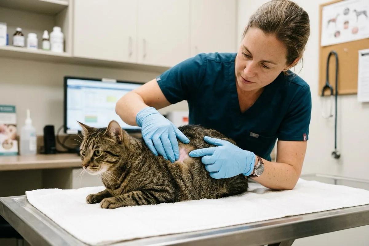
- Cutaneous Form: Typically solitary (or occasionally multiple) firm, well-circumscribed, raised, pinkish-white dermal nodules, most commonly located around the head, neck, and pinnae. In Siamese cats, a juvenile histiocytic variant can spontaneously regress.
- Splenic Form: Primary proliferation within the red pulp, causing massive organomegaly and displacing abdominal viscera.
- Intestinal Form: High-grade aggressive mural infiltration leading to mechanical obstruction and rapid regional lymph node metastases.
Systemic Signs and Histamine Degranulation
Neoplastic degranulation releases bioactive amines into circulation:
- Histamine-Induced Gastric Ulceration: Stimulation of parietal H2 receptors triggers hyperacidity, leading to inappetence, vomiting, hematemesis, and dark tarry melena.
- Heparin Release: Localized ecchymoses and prolonged bleeding tendencies.
- Darier's Sign: Transient erythema, edema, and intense pruritus following manipulation of the lesion.
- Visceral Manifestations: Weight loss, lethargy, marked anemia, and a palpable mid-abdominal mass.
Diagnostics and Staging
- Fine-Needle Aspiration Cytology (FNAC): Readily identifies discrete round cells with central nuclei and characteristic purple-magenta metachromatic granules.
- Peripheral Blood Smear Evaluation: Critical in splenic cases to detect circulating atypical mast cells (mastocytemia).
- Abdominal Ultrasonography: Demonstrates diffuse moth-eaten or swiss-cheese hypoechoic parenchymal mottling of the spleen.
- Histopathology: Differentiates compact well-differentiated tumors from pleomorphic high-grade variants.
Therapeutic Approaches
- Cutaneous Excision: Narrow surgical margins (0.5–1 cm) yield cure rates exceeding 85–90% for well-differentiated mastocytomas.
- Splenectomy for Splenic Disease: Complete surgical removal of the spleen provides significant relief from degranulation and median survival times of 12 to 18 months, even in cats with demonstrable pre-operative mastocytemia.
- Medical Supportive Therapy: H1 antihistamines (diphenhydramine) and H2 antagonists (famotidine), accompanied by receptor tyrosine kinase inhibitors (toceranib phosphate) for non-resectable or c-KIT mutated disease.
Prognosis
Well-differentiated cutaneous forms carry an excellent long-term prognosis following excision. Splenic mastocytoma responds favorably to surgical removal, whereas intestinal mastocytoma requires guarded monitoring.
Scientific sources and references
- Henry, C. et al. (2020). Feline mast cell tumors: a comprehensive review of diagnosis, staging, and therapeutic strategies. Journal of Feline Medicine and Surgery, 22(8), 731-744.
- Sabattini, S. et al. (2021). Histological grading of feline cutaneous mast cell tumors: validation of a two-tier system. Veterinary Pathology, 58(5), 903-912.
- Evans, C. et al. (2022). Outcome following splenectomy for splenic mast cell tumors in 52 cats: a retrospective multicenter study. Journal of Veterinary Internal Medicine, 36(3), 1089-1098.
Caring for a cat? Keep their health in your pocket
Register your cat on Nekolife to track vaccination history, weight, and get advice from loving cat parents.
- Vaccine & weight records
- Supportive cat community
- Earn Meow Points & badges
Related pages
Know someone who needs this?