Feline Pyothorax: Septic Pleural Empyema, Suffocation Signs and Treatment
Feline pyothorax (septic pleural empyema) is a life-threatening medical and surgical emergency marked by the accumulation of gross purulent exudate within the pleural space, compressing the lungs and precipitating severe hypoxemia. Frequently triggered by bite wounds inflicted during cat fights, migrating foreign bodies, or rupturing pulmonary parenchymal infections, pyothorax demands immediate hands-off oxygen supplementation, emergency therapeutic thoracocentesis, and bilateral chest tube placement with pleural lavage.
In this guide
Etiology and Pathogenesis of Pleural Empyema
The pleural space normally contains only a thin film of transudative fluid facilitating smooth pulmonary gliding. In pyothorax, pathogenic bacteria colonize this space, triggering severe inflammatory exudation:
- Bite wounds: Most frequent in outdoor or territorial cats. Puncture wounds inoculate normal feline oral flora (Pasteurella multocida, obligate anaerobes like Bacteroides, Fusobacterium, and Peptostreptococcus). The tiny skin punctures seal rapidly while anaerobic bacteria proliferate in the closed pleural cavity.
- Pneumonia and pulmonary abscesses: Underlying necrotizing pneumonia eroding through the visceral pleura into the pleural vault.
- Inhaled migrating plant awns: Foreign bodies that track through airways and penetrate the parenchyma.
- Hematogenous dissemination: Rare systemic bacteremia seeding the pleural lining.
Clinical Manifestations and Restrictive Dyspnea
Cats are notoriously adept at hiding chronic pleuropulmonary disease until acute respiratory collapse occurs:
- Restrictive breathing pattern: Rapid, shallow respirations with an exaggerated abdominal component (paradoxical breathing).
- Orthopneic posture: Sternally recumbent, neck extended, elbows abducted, and occasional open-mouth panting indicating critical decompensation.
- Muffled cardiopulmonary sounds: Ventral dullness upon percussion and faint heart sounds due to fluid shielding.
- Systemic signs: Lethargy, inappetence, dehydration, and either high fever or paradoxical hypothermia (a hallmark of septic shock in cats).
Emergency Stabilization and Diagnostic Protocol
Minimizing physical stress during initial triage is paramount to avoid fatal cardiac arrest:
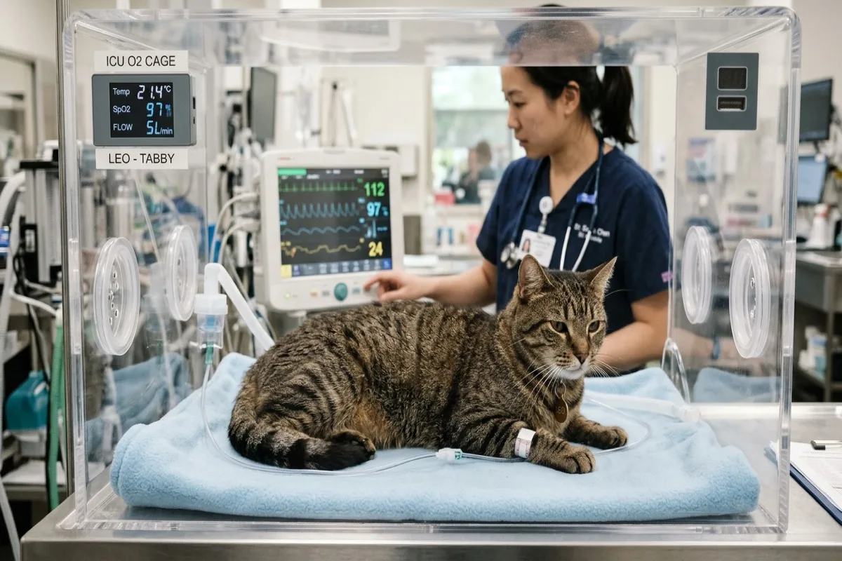
- Flow-by oxygen therapy: Pre-oxygenation in an oxygen chamber before any extensive diagnostic handling.
- Emergency needle thoracocentesis: Performed at the 7th–8th intercostal space at the costochondral junction. Aspiration of foul-smelling, opaque, tan-to-reddish exudate confirms diagnosis and provides life-saving pulmonary re-expansion.
- Cytology and microbiology: Microscopic evaluation demonstrates degenerate neutrophils and intracellular bacteria. Samples must be submitted for both aerobic and anaerobic cultures.
- Thoracic ultrasound (TFAST) and radiography: Obtained only after the patient has stabilized post-thoracocentesis to evaluate lung lobes and rule out masses.
Therapeutic Regimen: Chest Tubes, Lavage, and Antimicrobials
Repeated needle drainage alone yields poor survival rates. Standard veterinary protocol consists of:
- Bilateral thoracostomy tube placement: Enables continuous drainage and twice-daily warm sterile saline pleural lavages to evacuate thick fibrinous debris.
- Intravenous broad-spectrum antimicrobials: Empirical combination of ampicillin-sulbactam or amoxicillin-clavulanate alongside a fluoroquinolone or metronidazole, refined following culture results.
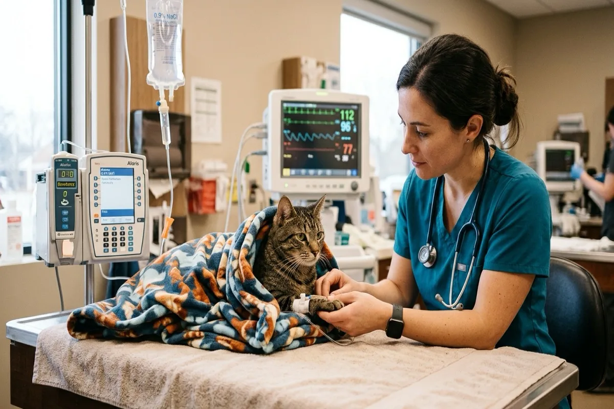
- Multimodal analgesia and fluid support: Balanced isotonic crystalloids and opioid analgesics (such as buprenorphine).
- Surgical exploration (thoracotomy/VATS): Indicated for non-resolving pockets, restrictive pleuritis, or foreign body retrieval.
Prognosis and Home Care
With timely stabilization and thorough tube drainage, survival rates range from 75% to 85%. Oral antibiotic therapy must be continued for at least 4 to 6 weeks following tube removal to eradicate residual micro-abscesses.
Scientific sources and references
- Boothe, H. W. et al. (2021). Feline pyothorax: clinical presentation, diagnosis, and long-term management. Journal of Feline Medicine and Surgery, 23(8), 712-724.
- Stillion, J. R., & Letendre, J. (2020). A review of feline pyothorax: etiology, diagnosis, treatment, and outcomes. Journal of Veterinary Emergency and Critical Care, 30(1), 17-29.
- MacPhail, C. (2022). Surgical management of thoracic emergencies in small animals. Veterinary Clinics of North America: Small Animal Practice, 52(2), 385-398.
Caring for a cat? Keep their health in your pocket
Register your cat on Nekolife to track vaccination history, weight, and get advice from loving cat parents.
- Vaccine & weight records
- Supportive cat community
- Earn Meow Points & badges
Related pages

Know someone who needs this?