Feline Septic Peritonitis: Organ Perforation, Hypothermia and Emergency Surgery
Feline septic peritonitis is one of the most catastrophic medical and surgical emergencies in feline practice, defined by overwhelming bacterial inflammation of the peritoneal cavity resulting from full-thickness rupture of a hollow abdominal organ (linear gastrointestinal foreign bodies, ulcer perforation, dehiscence of a surgical anastomosis, or rupture of an infected biliary/urinary tract). Cats with sepsis display a deceptively muted hemodynamic profile characterized by the "feline septic shock triad" (hypothermia, relative bradycardia, and profound hypotension), requiring emergency ultrasound-guided abdominocentesis and immediate surgical exploratory laparotomy.
In this guide
Etiology and Visceral Sources of Contamination
- Gastrointestinal Perforation (> 60% of cases): Linear foreign bodies (string/yarn sawing through mesenteric borders), penetrating bones or needles; perforating ulcerations associated with gastrointestinal lymphoma or corticosteroid/NSAID toxicity; surgical wound dehiscence following enterectomy (typically days 3–5).
- Biliary and Urogenital Tract Rupture: Necrotizing cholecystitis leading to septic bile peritonitis; ruptured pyometra in intact queens or prostatic abscessation.
- Penetrating External Trauma: Deep animal bite wounds or abdominal punctures.
The Feline Septic Shock Triad
Cats deviate sharply from the hyperdynamic, febrile septic shock seen in dogs and humans:
- Hypothermia, Bradycardia, and Hypotension: Severe core hypothermia (< 98.5 °F / 37.0 °C) accompanied by relative bradycardia (< 140 bpm) and collapsed peripheral vascular tone.
- Acute Abdomen: Guarding, tucked hunched posture, vocalization upon light abdominal palpation, or recumbent apathy.
- Poor Perfusion: Pale or muddy gray mucous membranes, capillary refill time > 2.5 seconds, and weak femoral pulses.
Diagnosis via Abdominocentesis
Prompt abdominocentesis provides rapid definitive confirmation:
- Abdominal FAST (AFAST): Rapidly detects free peritoneal fluid in dependent pockets.
- Effusion Cytology: Demonstration of intracellular bacteria within degenerating, vacuolated neutrophils is pathognomonic for septic peritonitis.
- Biochemical Blood-to-Fluid Gradients:
- Glucose Difference: Peritoneal fluid glucose > 20 mg/dL lower than concurrent peripheral blood glucose due to rapid microbial and leukocytic consumption.
- Lactate Difference: Peritoneal fluid lactate > 2.0 mmol/L higher than peripheral blood lactate.
Emergency Resuscitation and Surgical Management
Septic peritonitis caused by visceral leakage is an absolute surgical condition; antibiotics alone cannot heal an active perforation:
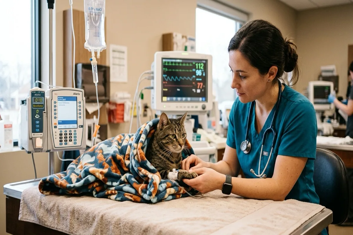
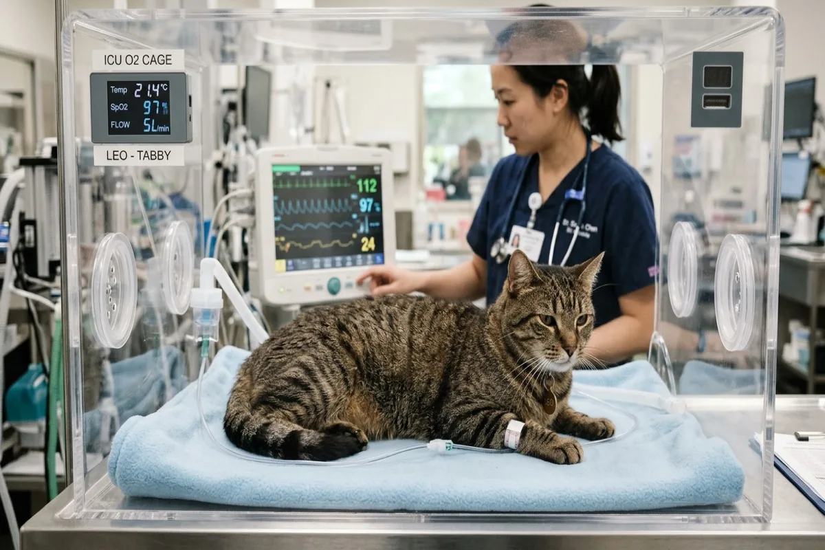
- Aggressive Pre-operative Resuscitation: Balanced warmed intravenous crystalloids, vasopressor therapy (norepinephrine/dopamine) for persistent hypotension, and warming devices.
- Broad-Spectrum Parenteral Antibiotics: Immediate IV administration of ampicillin-sulbactam combined with metronidazole and a fluoroquinolone (e.g., marbofloxacin).
- Emergency Exploratory Laparotomy: Identification of the perforation site, resection and anastomosis (enterectomy) or primary closure with an omental patch, copious peritoneal lavage with warm sterile saline (liters of lavage), and placement of a closed-suction drain (Jackson-Pratt).
- Early Enteral Nutrition: Placement of an esophagostomy or gastrostomy feeding tube before abdominal closure.
Prognosis
Historically mortality rates range between 30% and 50%. However, with rapid resuscitation, aggressive surgical debridement, and dedicated intensive care, survival rates of 60% to 70% are achievable.
Scientific sources and references
- Costello, M. F. et al. (2020). Septic peritonitis in cats: 76 cases (2009-2019). Journal of Veterinary Emergency and Critical Care, 30(4), 438-447.
- Bonczynski, J. J. et al. (2021). Comparison of peritoneal fluid and peripheral blood glucose and lactate concentrations for the diagnosis of septic peritonitis in cats. Journal of the American Veterinary Medical Association, 259(1), 54-61.
- MacPhail, C. (2022). Peritonitis and abdominal sepsis in small animals. Veterinary Clinics of North America: Small Animal Practice, 52(2), 401-417.
Caring for a cat? Keep their health in your pocket
Register your cat on Nekolife to track vaccination history, weight, and get advice from loving cat parents.
- Vaccine & weight records
- Supportive cat community
- Earn Meow Points & badges
Related pages
Know someone who needs this?