Uveitis in Cats: Ocular Signs, Aqueous Flare, and Secondary Glaucoma Risks
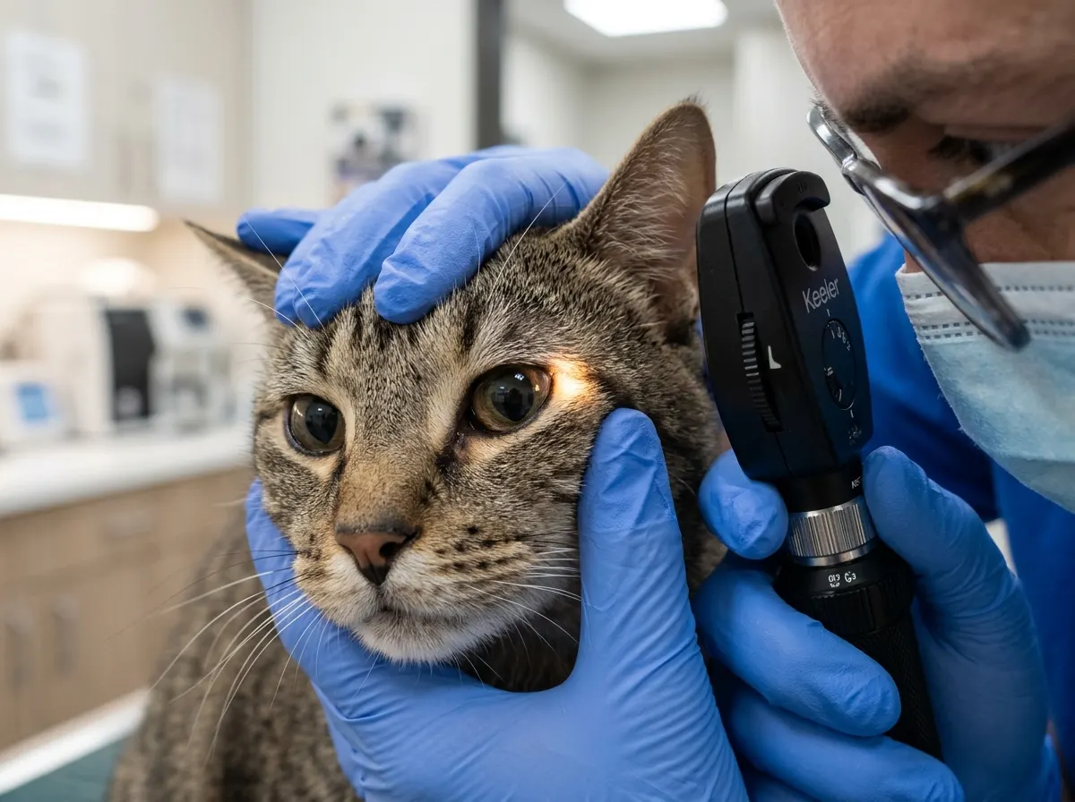
Feline uveitis is inflammation of the uveal tract—the vascular tunic of the eye encompassing the iris, ciliary body, and choroid. In feline medicine, uveitis is seldom a benign local phenomenon; in the vast majority of cases, it represents the primary ocular hallmark of a life-threatening systemic infectious, inflammatory, or neoplastic disorder (such as Feline Infectious Peritonitis [FIP], Feline Leukemia Virus [FeLV], Feline Immunodeficiency Virus [FIV], systemic toxoplasmosis, or lymphoma). Without prompt intervention, breakdown of the blood-aqueous barrier results in posterior synechiae, cataracts, and secondary glaucoma leading to irreversible blindness.
In this guide
Clinical Hallmarks and Slit-Lamp Findings
- Aqueous Flare (Tyndall Effect): Cloudiness of the anterior chamber caused by protein and cellular leakage through inflamed ciliary vasculature when examined with a narrow slit-lamp beam.
- Hypopyon and Hyphema: Gravitational pooling of inflammatory fibrinous exudate (hypopyon) or free erythrocytes (hyphema) in the ventral anterior chamber.
- Spastic Miosis: A constricted, non-responsive pupil caused by inflammatory prostaglandins irritating the iris sphincter muscle.
- Keratic Precipitates (KPs): Distinct cellular inflammatory clusters adhering to the posterior corneal endothelium.
- Ocular Hypotony: Significantly decreased intraocular pressure (IOP often < 10 mmHg) due to compromised aqueous humor secretion.
Diagnostic Protocol
Every case requires a systemic investigation: FeLV/FIV serology, Toxoplasma gondii titers, FIP evaluation (A:G ratio, RT-PCR), serial tonometry, and thoracic/abdominal imaging to rule out multicentric lymphoma. Medical therapy combines topical corticosteroids (prednisolone acetate, only if corneal ulceration is ruled out via fluorescein test), 1% atropine sulfate to achieve cycloplegia and prevent synechiae, and targeted systemic therapy (GS-441524 for FIP, clindamycin for toxoplasmosis).
Scientific Sources and References
- Stiles, J. (2014). Feline ophthalmology. In Gelatt, K. N. (Ed.), Essentials of Veterinary Ophthalmology (3rd ed., pp. 411-450). Wiley-Blackwell.
- Townsend, W. M. (2008). Feline uveitis: feline leukemia virus, feline immunodeficiency virus, and feline infectious peritonitis. Veterinary Clinics of North America: Small Animal Practice, 38(2), 365-378.
Caring for a cat? Keep their health in your pocket
Register your cat on Nekolife to track vaccination history, weight, and get advice from loving cat parents.
- Vaccine & weight records
- Supportive cat community
- Earn Meow Points & badges
Related pages
Know someone who needs this?