Feline Triaditis: Diagnosing Concurrent Cholangitis, Pancreatitis and Bowel Disease
Feline triaditis is a complex inflammatory syndrome unique to domestic cats, characterized by concurrent and simultaneous inflammation across three critical abdominal systems: the hepatobiliary tree (cholangitis/cholangiohepatitis), the pancreas (acute or chronic pancreatitis), and the gastrointestinal tract (inflammatory bowel disease or IBD). This multi-organ convergence is directly tied to feline anatomy, wherein the common bile duct and the primary pancreatic duct unite into a single shared channel before emptying into the duodenum, creating a vulnerable pathway for biliary-pancreatic reflux and ascending bacterial migration.
In this guide
Pathophysiology and Anatomical Predispositions
Unlike canine and human anatomy, over 80% of felines lack an accessory pancreatic duct. All exocrine pancreatic secretions enter the duodenal lumen via the major pancreatic duct (duct of Wirsung), which fuses with the common bile duct at the major duodenal papilla.
Additionally, the feline duodenum naturally supports an intestinal bacterial load up to one hundred times denser than that of canines. Consequently, any underlying condition causing duodenal vomiting, increased intraluminal pressure, or mucosal barrier disruption promotes reverse flow of digestive enzymes and enteric bacteria into both the pancreatic parenchyma and the biliary tree, sparking self-amplifying tissue autodigestion and infection.
Clinical Signs and Warning Flags
The clinical manifestation of feline triaditis is often subtle, insidious, and non-specific:
- Severe lethargy and social withdrawal: The cat adopts a hunched, guarded posture, hiding in dark quiet spots and resisting handling.
- Anorexia and persistent hyporexia: Fasting for more than 48 hours carries a severe risk of secondary feline hepatic lipidosis.
- Intermittent vomiting: Regurgitation of clear fluid, yellow bile, or thick gastric mucus.
- Overt jaundice (icterus): Yellowish discoloration of the sclera, ear pinnae, and oral mucosa caused by hyperbilirubinemia.
- Cranial abdominal discomfort: Intense abdominal splinting and vocalization upon palpation due to peripancreatic fat necrosis and localized peritonitis.
- Weight loss and chronic soft stools: Poor intestinal absorption and exocrine dysfunction leading to steatorrhea and muscle wasting.
Diagnostic Workup
Definitive confirmation relies on a comprehensive diagnostic panel:
- Complete Biochemistry & Liver Panel: Significant elevation in liver enzymes (ALT, AST, ALP, GGT) along with hyperbilirubinemia.
- Feline Pancreatic Lipase Immunoreactivity (Spec fPL / fPLI): The gold standard serum test to detect active pancreatic necrosis and inflammation.
- Cobalamin (Vitamin B12) & Folate Levels: Essential for staging chronic ileal and jejunal malabsorption related to concurrent IBD.
- High-Resolution Abdominal Ultrasonography: Crucial for visualizing duodenal wall thickening, hypoechoic pancreatic swelling with hyperechoic mesenteric fat halo, and thickened gall bladder walls with biliary sludge.
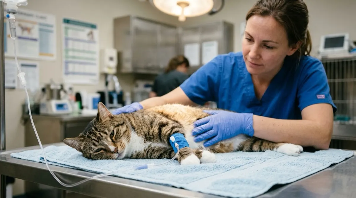
Inpatient Hospitalization and Critical Care
Effective management requires immediate inpatient stabilization:
- Balanced Intravenous Fluid Therapy: Crystalloid therapy supplemented with potassium chloride to correct dehydration, electrolyte derangements, and optimize pancreatic microcirculation.
- Early Multimodal Pain Management: Pancreatic visceral pain is agonizing in felines; pure or partial mu-opioids (buprenorphine, methadone) are mandatory.
- Antiemetic and Gastromucosal Support: Intravenous maropitant to suppress central and peripheral nausea, combined with ondansetron if refractory.
- Nutritional Support via Feeding Tube: Placement of an esophagostomy or nasoesophageal tube within 24 to 36 hours of anorexia is vital to halt hepatic steatosis.
- Targeted Antimicrobial Regimen: Prescribed whenever suppurative bacterial cholangitis is suspected, utilizing broad-spectrum antibiotics with good biliary excretion (amoxicillin-clavulanic acid or pradofloxacin).
Scientific Sources and References
- Simpson, K. W. (2015). Pancreatitis and triaditis in cats: clinical perspectives. Journal of Feline Medicine and Surgery, 17(9), 706-716.
- Fragkou, F. C., et al. (2016). Prevalence and clinicopathological features of triaditis in a prospective cohort of cats with inflammatory bowel disease. Journal of Veterinary Internal Medicine, 30(4), 1031-1045.
- Center, S. A. (2009). Feline cholangitis/cholangiohepatitis complex. Compendium on Continuing Education for the Practicing Veterinarian, 31(4), 166-176.
Caring for a cat? Keep their health in your pocket
Register your cat on Nekolife to track vaccination history, weight, and get advice from loving cat parents.
- Vaccine & weight records
- Supportive cat community
- Earn Meow Points & badges
Related pages
Know someone who needs this?